Neuronal cells may be repaired in the near future opening the potential to reverse brain damage, spinal cord injuries and the devastating effects of diseases such as MS, ALS, Parkinson’s, Dementia and Alzheimer. For the first time researchers have direct evidence that viruses can infect neuronal cells, raising hope that antiviral therapy might be effective against neurologic diseases and cell damage, according to a new study.
A neurological disorder is any disorder of the body nervous system. Structural, biochemical or electrical abnormalities in the brain, spinal cord or other nerves can result in a range of symptoms. Examples of symptoms include paralysis, muscle weakness, poor coordination, loss of mental control, sensation, seizures, confusion, pain and altered levels of consciousness
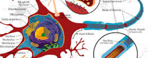
Neurons are nerve cells that transmit nerve signals to and from the brain at up to 200 mph. The neuron consists of a cell body (or soma) with branching dendrites (signal receivers) and a projection called an axon, which conduct the nerve signal. At the other end of the axon, the axon terminals transmit the electro-chemical signal across a synapse (the gap between the axon terminal and the receiving cell). The word “neuron” was coined by the German scientist Heinrich Wilhelm Gottfried von Waldeyer-Hartz in 1891 (he also coined the term “chromosome”).
There are different types of neuronal cells. They all carry electro-chemical nerve signals, but differ in structure (the number of processes, or axons, emanating from the cell body) and are found in different parts of the body.
- Sensory neurons or Bipolar neurons carry messages from the body’s sense receptors (eyes, ears, etc.) to the CNS. These neurons have two processes. Sensory neuron account for 0.9% of all neurons. (Examples are retinal cells, olfactory epithelium cells.)
- Motoneurons or Multipolar neurons carry signals from the CNS to the muscles and glands. These neurons have many processes originating from the cell body. Motoneurons account for 9% of all neurons. (Examples are spinal motor neurons, pyramidal neurons, Purkinje cells.)
- Interneurons or Pseudopolare (Spelling) cells form all the neural wiring within the CNS. These have two axons (instead of an axon and a dendrite). One axon communicates with the spinal cord; one with either the skin or muscle. These neurons have two processes. (Examples are dorsal root ganglia cells.)
The core component of the nervous system in general, and the brain in particular, is the neuron or nerve cell, the “brain cells” of popular language. A neuron is an electrically excitable cell that processes and transmits information by electro-chemical signalling. Unlike other cells, neurons never divide, and neither do they die off to be replaced by new ones. By the same token, they usually cannot be replaced after being lost, although there are a few exceptions.
Information transmission within the brain, such as takes place during the processes of memory encoding and retrieval, is achieved using a combination of chemicals and electricity. It is a very complex process involving a variety of interrelated steps, but a quick overview can be given here.
Unlike other body cells, most neurons in the human brain are only able to divide to make new cells (a process called neurogenesis) during fetal development and for a few months after birth.
These brain cells may increase in size until the age of about eighteen years, but they are essentially designed to last a lifetime.
During childhood, and particularly during adolescence, a process known as “synaptic pruning” occurs.
Although the brain continues to grow and develop, the overall number of neurons and synapses are reduced by up to 50%, removing unnecessary neuronal structures and allowing them to be replaced by more complex and efficient structures, more suited to the demands of adulthood.
The data demonstrate that Epstein Barr Virus (EBV) and Kaposi’s sarcoma–associated herpesvirus (KSHV) are able to effectively infect neuronal cell lines, as well as primary neurons. “This phenomenon may potentially provide clues to their contribution to the pathogenesis of human neural diseases,” stated the researchers, led by senior author Erle S. Robertson, PhD, of the University of Pennsylvania in Philadelphia, PA.
“In this study, for the first time, we have successfully demonstrated the in vitro infection of Sh-Sy5y (a human neuroblastoma cell line) and Ntera2 cells (a neuronally committed human teratocarcinoma cell line), as well as human primary neurons. We have also determined that the infection is predominately lytic. Additionally, we also report infection of neuronal cells by KSHV in vitro similar to that by EBV,” stated the authors.
They believe that these findings may “open new avenues of consideration related to neuronal pathologies and infection with these viruses. Furthermore, their contribution to chronic infection linked to neuronal disease will provide new clues to potential new therapies.”
In many debilitating neurodegenerative diseases, including MS, the efficiency of electrochemical signals is greatly reduced, they noted, often leading to problems related to cognition and muscular activity. “It is likely that viral infection with EBV or KSHV may severely reduce these signals, leading to reduced cognition and reduced neuromuscular function,” they stated.
The results clearly show that infection of neuronal cells can occur, but further studies are needed to understand the underlying mechanism of these infections and their relevance to neuronal diseases.
Sources:
http://www.neurologytimes.com/alzheimer-disease/herpes-viruses-infection-neurons#sthash.kOMeJYI8.dpuf
http://www.human-memory.net/brain_neurons.html
http://www.enchantedlearning.com/subjects/anatomy/brain/Neuron.shtml